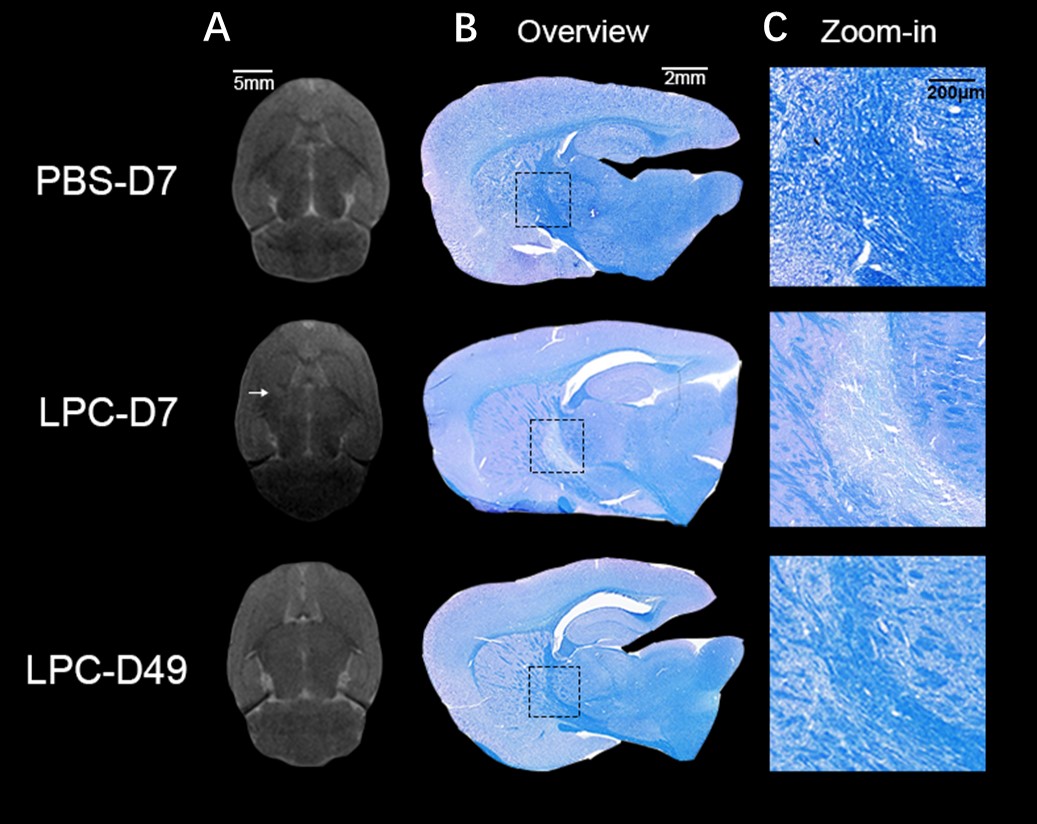
In vivoand histological observations of demyelination and remyelination time points in the model. (A) T2 weighted MRI structural scan results. Arrow indicates higher signal intensity compared to the normal internal capsule. (B) LFB-eosin myelin staining of frozen sections near the lesion site shows lighter staining at the internal capsule in the LPC-D7 group, while LPC-D49 returned normal. (C) Zoom-ins are all magnified images from the dashed box.